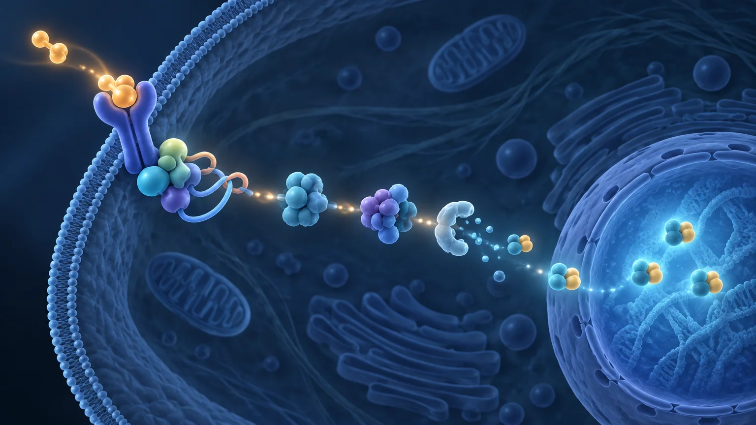
TLDR: The TNF alpha NF-kB signaling pathway begins when TNF-α engages TNFR1 and promotes formation of a membrane-associated signaling complex. Adaptor proteins and nondegradative ubiquitin chains organize the recruitment of TAK1 and the IKK complex. Activated IKK phosphorylates IκBα, leading to its ubiquitination and proteasomal degradation. Freed from IκBα restraint, canonical NF-κB—commonly the RelA/p65:p50 dimer—accumulates in the nucleus and regulates transcription. Newly produced IκBα then contributes to negative feedback.
The central idea is simple, but the mechanism is not a single chain of proteins passing a signal from one to the next. The TNF alpha NF-kB signaling pathway is better understood as a dynamically assembled, ubiquitin-organized network. It can promote NF-κB-dependent transcription, connect with MAPK signaling, or contribute to cell-death-associated processes depending on the molecular and cellular context. A pathway diagram therefore describes one major route through the network, not an inevitable outcome for every TNF-stimulated cell.
The TNF alpha NF-kB signaling pathway at a glance
For the canonical TNFR1-centered pathway, the core sequence is: TNF-α engagement of TNFR1; assembly of receptor complex I; recruitment of TRADD, RIPK1, TRAF proteins, cIAP proteins, and LUBAC; construction of ubiquitin signaling scaffolds; recruitment and activation of TAK1 and the NEMO-containing IKK complex; phosphorylation and degradation of IκBα; and nuclear accumulation of NF-κB.
| Stage | Principal components | Functional consequence |
|---|---|---|
| Receptor engagement | TNF-α and TNFR1 | Creates a receptor-associated platform for signaling-complex assembly |
| Complex I assembly | TRADD, RIPK1, TRAF2/5, cIAP1/2, LUBAC | Organizes receptor-proximal signaling and ubiquitin scaffolds |
| Kinase recruitment | TAK1 module and IKK complex | Connects ubiquitin-dependent organization to kinase activation |
| Inhibitor removal | IKK and IκBα | Phosphorylated IκBα is ubiquitinated and degraded |
| Nuclear response | RelA/p65:p50 and other NF-κB dimers | NF-κB accumulates in the nucleus and regulates responsive genes |
| Feedback | Newly synthesized IκBα and other regulators | Limits or reshapes the amplitude and duration of signaling |
Step 1: TNF-α engages TNFR1
TNF-α can signal through TNFR1 and TNFR2, but these receptors should not be treated as interchangeable. The widely taught rapid canonical NF-κB route is usually framed around TNFR1 because it provides the receptor-proximal architecture described here. TNFR2 uses a different pattern of adaptor engagement and can produce context-dependent signaling that falls outside this simplified TNFR1 sequence.
TNF-α binding changes the organization of TNFR1 and enables cytoplasmic signaling proteins to assemble at the receptor. This initial membrane-associated assembly is commonly called TNFR1 complex I. The word “complex” matters: receptor engagement does not directly activate NF-κB. It creates a temporary molecular platform on which adaptors, ubiquitin ligases, ubiquitin chains, and kinase modules can be organized.
Step 2: TNFR1 complex I recruits adaptor proteins
TRADD and RIPK1 help organize the receptor-proximal complex, while TRAF2 or TRAF5 and the cellular inhibitor-of-apoptosis proteins cIAP1 and cIAP2 support downstream signaling. LUBAC, the linear ubiquitin chain assembly complex, also becomes part of this functional network. The exact abundance, order, and persistence of these components can vary with cell type, time after stimulation, protein expression, and regulatory modifications.
These proteins do not all perform the same job. TRADD is principally an adaptor in this setting. RIPK1 can act as a scaffold, although it also has kinase-dependent functions in other TNFR1 outcomes. TRAF and cIAP proteins help organize ubiquitin-dependent signaling, while LUBAC generates M1-linked, also called linear, ubiquitin chains. Collapsing these roles into a generic label such as “activators” hides the biochemical organization that makes the pathway work.
Step 3: Ubiquitin chains build signaling scaffolds
Ubiquitin is often introduced as a tag that sends proteins to the proteasome. That is only one of its functions. In TNFR1 signaling, particular ubiquitin-chain architectures serve as nondegradative docking surfaces that recruit and stabilize downstream signaling assemblies. K63-linked chains are associated with recruitment of the TAB2/TAB3–TAK1 module, while LUBAC-generated M1-linked chains support engagement of NEMO and the IKK complex.
This creates an important distinction: ubiquitination near TNFR1 does not necessarily mean that the modified receptor-complex protein is about to be destroyed. In this phase, ubiquitin can encode molecular organization. By bringing kinase modules into proximity, the chains help convert receptor engagement into coordinated enzymatic activity.
The topology is also regulated rather than permanent. Chain formation, recognition, editing, and removal influence whether complex I remains competent for transcriptional signaling. A static pathway illustration cannot show all of these competing reactions, so it should be read as an orientation map rather than a complete molecular inventory.
Step 4: TAK1 and the IKK complex relay the signal
The TAK1 kinase module and the IKK complex are recruited through interactions supported by the ubiquitin-rich signaling platform. TAK1-dependent signaling contributes to activation of the canonical IKK complex in the TNFR1 pathway. The core IKK complex contains two related catalytic subunits, IKKα and IKKβ, plus the regulatory scaffold NEMO, also called IKKγ.
IKKβ has a particularly prominent role in phosphorylation of canonical IκB substrates, but the complex should not be reduced to IKKβ alone. IKKα participates in the complex, and NEMO helps the kinase machinery recognize and respond to ubiquitin-organized signaling. Readers looking for a broader pathway overview can consult this review of NF-κB signaling and its regulatory modules.
Step 5: IKK marks IκBα for removal
In an unstimulated cell, IκB proteins restrain NF-κB dimers, including by limiting their nuclear accumulation. Following TNFR1 signaling, activated IKK phosphorylates IκBα at regulatory sites. This phosphorylation licenses subsequent ubiquitination of IκBα and its degradation by the proteasome.
The identity of the degraded protein is a frequent source of confusion. IκBα is the inhibitor being removed; NF-κB itself is not destroyed during this activation step. Degradation of IκBα exposes or makes available the nuclear-localization behavior of the associated NF-κB dimer, allowing the transcription factor to accumulate in the nucleus.
IκB and IKK are different protein systems
Their similar names can obscure distinct functions. IκBα is an inhibitory binding protein that restrains NF-κB. IKK is the kinase complex that phosphorylates IκBα. In concise terms, IKK acts on IκBα; loss of IκBα releases NF-κB. Keeping those three entities separate prevents a common but consequential pathway error.
- IKKα and IKKβ are catalytic kinase subunits.
- NEMO, or IKKγ, is the regulatory component of the canonical IKK complex.
- IκBα binds and restrains NF-κB before stimulation.
- Phosphorylated IκBα is ubiquitinated and degraded by the proteasome.
- NF-κB can then accumulate in the nucleus and affect transcription.
Step 6: RelA:p50 accumulates in the nucleus
RelA, also called p65, paired with p50 is a common NF-κB dimer in canonical signaling. After IκBα loss, this dimer can accumulate in the nucleus, bind appropriate regulatory DNA sequences, interact with transcriptional machinery, and alter gene expression. A closer treatment of the subunits and their division of labor is available in the guide to the NF-κB p50 and p65 heterodimer.
Nuclear localization is an important measurement, but it is not equivalent to every downstream conclusion. Detecting nuclear RelA supports pathway activation more directly than a generic viability change, yet it does not by itself identify which genes were transcribed, whether a particular target protein changed, or whether the resulting phenotype depends on NF-κB. Those questions require additional measurements.
Step 7: Negative feedback limits and reshapes the response
NF-κB activation is not simply an irreversible switch. NF-κB-dependent transcription includes negative-feedback regulators, notably newly synthesized IκBα. New IκBα can enter the nucleus, bind NF-κB, and help reduce or redistribute its transcriptional activity. This feedback contributes to the amplitude and duration of the response and can support dynamic behavior rather than a sustained maximum signal.
Consequently, two experiments conducted at different sampling times can appear to disagree even when both are technically sound. An early time point may capture IκBα loss and nuclear RelA accumulation, whereas a later measurement may capture IκBα re-expression and reduced nuclear signal. A single endpoint can therefore miss the trajectory of pathway activation.
TNFR1 can signal beyond canonical NF-κB
TNFR1 complex I is associated with transcriptional pathways that include NF-κB and MAPK signaling. Under different molecular conditions, TNFR1-associated components can reorganize into cell-death-related complexes connected with apoptosis or necroptosis. These are branches of a regulated network, not automatic consequences of TNF exposure.
RIPK1 illustrates why pathway labels must be used carefully. Its scaffold function in receptor-proximal signaling is conceptually distinct from kinase-dependent roles associated with certain death-signaling contexts. Likewise, reduced cell viability after TNF exposure does not, by itself, demonstrate apoptosis, necroptosis, or failure of NF-κB signaling. The mechanism must be established with branch-appropriate controls and readouts.
Canonical and noncanonical NF-κB are not the same pathway
The TNFR1 route described above is canonical NF-κB signaling: it uses a NEMO-containing IKK complex, rapidly removes inhibitory IκB proteins, and commonly mobilizes RelA:p50. The noncanonical pathway instead centers on NIK-dependent activation of IKKα and processing of the p100 precursor to p52, commonly supporting RelB:p52 activity. It is engaged by a more restricted set of receptors and follows different regulatory logic.
TNF receptor family membership therefore does not mean every receptor activates NF-κB through an identical mechanism. When interpreting an experiment, receptor identity, adaptor usage, NF-κB dimer composition, and the measured time course should all be specified.
How to test the pathway experimentally
No single assay establishes every step from receptor engagement to transcriptional function. A stronger experimental design uses measurements matched to successive mechanistic questions.
- Receptor-proximal assembly: use interaction or complex-isolation methods to ask whether expected adaptors associate with TNFR1 after stimulation.
- Ubiquitin organization: measure relevant ubiquitin modifications while controlling for linkage specificity and the purity of isolated complexes.
- Kinase activation: assess TAK1 and IKK activation with appropriate phosphorylation or activity measurements rather than inferring kinase activity solely from a downstream phenotype.
- IκBα removal: follow phosphorylation and total IκBα abundance over a time course. Proteasome inhibition can help test whether loss depends on proteasomal degradation, although inhibitors can have broader cellular effects.
- Nuclear accumulation: use imaging, cell fractionation, or related methods to measure RelA localization. Fractionation requires controls for cytoplasmic and nuclear contamination.
- DNA association and transcription: measure promoter or enhancer occupancy where relevant, then examine transcript changes. Nuclear localization alone does not prove regulation of a specific gene.
- Functional dependence: perturb a defined pathway component and test whether the molecular or cellular phenotype changes. Include controls that distinguish pathway-specific effects from nonspecific toxicity.
Cell type, TNF exposure, sampling time, basal protein abundance, and post-translational regulation can all alter the observed response. Single-cell studies also show that population averages can conceal heterogeneous signaling and fate decisions. Replication across models strengthens generalizability, but it does not erase biological differences among those models.
What the pathway does—and does not—establish
TNF and NF-κB signaling are studied in inflammatory biology, immune regulation, cancer research, and cell-death mechanisms. Those associations make the pathway biologically important, but they do not turn every pathway component into a validated therapeutic target. A biochemical interaction, a cell-culture phenotype, an animal-model result, and a human clinical outcome are different levels of evidence.
The strongest interpretation should stay close to the experiment. IκBα loss supports activation of the canonical signaling machinery. Nuclear RelA supports nuclear pathway engagement. A transcript change supports a downstream expression response. A disease-relevant phenotype requires additional causal evidence, and a preclinical result does not establish clinical benefit or treatment guidance.
Conclusion
The most useful way to understand TNF-α-driven canonical NF-κB signaling is as an ordered but adaptable assembly process. TNFR1 recruits complex I; adaptor proteins and ubiquitin chains construct a signaling platform; TAK1 and IKK relay the signal; IκBα is phosphorylated, ubiquitinated, and degraded; and NF-κB accumulates in the nucleus. Feedback then reshapes the response, while MAPK and cell-death-related branches remain possible in the appropriate context.
When reading or designing an experiment, identify the exact stage being measured. Receptor association, IκBα degradation, nuclear localization, target-gene expression, and cellular phenotype answer different questions. Connecting those levels with time-resolved measurements and appropriate perturbations is more informative than treating a single readout as proof of the entire pathway.
References
- Cellular heterogeneity in TNF/TNFR1 signalling: live cell imaging of cell fate decisions in single cells – PMC
- More to life than NF-κB in TNFR1 signaling – PMC
- Reactome | Membrane-anchored TNF-α binds TNFR1 (R-HSA-83660)
- Tumor Necrosis Factor Receptor-Associated Factor Regulation of Nuclear Factor κB and Mitogen-Activated Protein Kinase Pathways – PMC
- NF-κB signaling – PMC
- Regulation of NF-κB by TNF Family Cytokines – PMC
- Tumor necrosis factor alpha-induced phosphorylation of I kappa B alpha is a signal for its degradation but not dissociation from NF-kappa B.
- Tumor necrosis factor and interleukin-1 lead to phosphorylation and loss of I kappa B alpha: a mechanism for NF-kappa B activation – PubMed
- IKK-1 and IKK-2: cytokine-activated IkappaB kinases essential for NF-kappaB activation – PubMed