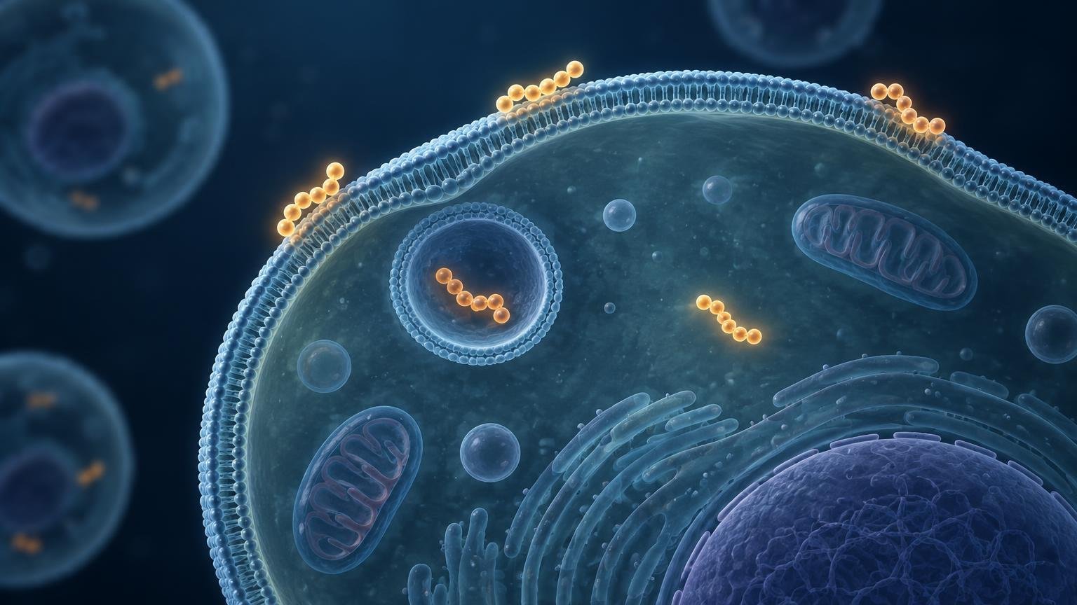
TLDR: A cell-associated cell-penetrating peptide signal is not automatically an intracellular signal. It can include peptide bound to the outer plasma membrane, material inside endosomes, and—in some cases—a much smaller cytosolically available fraction. A defensible experiment combines a surface-accessibility test with spatial imaging and, when cytosolic delivery matters, a cytosol-specific functional or biochemical assay. No single wash, quencher, image, or flow-cytometry measurement resolves every compartment.
The central problem in cell surface binding vs internalization cpp experiments is that many common assays measure total signal associated with a cell. Strong electrostatic binding to the plasma membrane can therefore look like efficient uptake, particularly when fluorescence is quantified without a control for extracellularly accessible label. Published CPP studies have explicitly separated membrane-bound and internalized populations, showing that membrane affinity does not necessarily predict internalization efficiency.
The early answer is to define the endpoint before selecting the assay. If the question is whether a CPP contacts cells, total association may be sufficient. If the question is whether it crosses the plasma membrane, surface-accessible material must be excluded. If the intended endpoint is intracellular target engagement, even true internalization is not enough: peptide trapped in endosomes has not necessarily reached a cytosolic target.
Three endpoints that should not be combined
| Endpoint | What it means | What it does not establish |
|---|---|---|
| Cell-surface association | CPP or cargo remains accessible from outside the cell, including material bound to membrane components | Internalization, endosomal entry, or cytosolic availability |
| Internalization | CPP or cargo is enclosed by the plasma membrane and no longer freely accessible from the extracellular medium | Escape from endosomes, target engagement, or biological activity |
| Cytosolic delivery | CPP or released cargo reaches the cytosolic space in a form detectable by an appropriate assay | Correct subcellular targeting, molecular function, or a therapeutic effect |
These categories represent an evidence hierarchy rather than interchangeable definitions of “uptake.” Internalized CPP can remain in endosomal or lysosomal compartments, making cytosolic delivery a stricter endpoint than entry into the cell. Nuclear delivery, when required, is another separate step.
Why cell-associated fluorescence can be misleading
Many CPPs are rich in positively charged residues. Their interaction with negatively charged components at the cell surface can produce persistent membrane association that survives ordinary buffer washes. A flow cytometer then records fluorescence per cell but does not inherently know whether each fluorescent molecule is outside the plasma membrane, inside a vesicle, or free in the cytosol.
The label itself can also change the experiment. A fluorophore may alter CPP hydrophobicity, charge, aggregation, membrane affinity, or trafficking. Its fluorescence can vary with local environment, pH, quenching, or degradation. Fluorescence-based comparisons consequently require controls for dye behavior as well as peptide behavior. Reviews and quantitative comparisons of CPP uptake methods describe these pitfalls and show why fluorescence and chemical quantification should be treated as complementary measurements. A quantitative comparison of fluorescence, flow cytometry, and mass-spectrometric CPP measurements provides a useful methods-level treatment.
Sample preparation introduces another risk. Redistribution during fixation has been reported in protein-transduction-domain and CPP experiments. Live-cell imaging is preferable when practical because it avoids treating a fixation-dependent distribution as the native localization pattern. Fixed-cell experiments are still usable, but fixation, permeabilization, and image-processing conditions need validation.
Methods for separating surface binding from internalization
Live-cell microscopy
Microscopy supplies spatial information that bulk fluorescence cannot. A membrane outline suggests surface association, while intracellular puncta are consistent with vesicular uptake. Colocalization with validated endosomal or lysosomal markers can strengthen the interpretation that the CPP occupies those compartments.
However, an optical section is not automatically proof of cytosolic access. Signal above or below the cell can overlap its projected area, and limited resolution can make closely apposed membrane signal look intracellular. Diffuse fluorescence may suggest cytosolic distribution, but background, released dye, detector saturation, and out-of-focus light can create a similar appearance. Z-stacks, orthogonal views, appropriate membrane markers, live imaging, and restrained display settings improve the analysis.
Extracellular fluorescence quenching
A membrane-impermeant quencher can suppress fluorescence from labels that remain accessible outside the cell. The residual, quencher-resistant signal is operationally classified as protected from the extracellular quencher and can be compared with total cell-associated signal. This approach is especially useful with flow cytometry because it adds a surface-accessibility control to a high-throughput single-cell measurement.
The assay must be matched to the fluorescent label. Trypan blue is commonly used as an extracellular fluorescence quencher, but its effectiveness depends on the fluorophore and measurement settings. Dithionite can chemically reduce and extinguish certain exposed fluorophores, but only when the label has suitable chemistry. Neither reagent should be assumed to work universally.
Quencher resistance means that the label was inaccessible to the quencher under the test conditions. It does not reveal whether the signal is in an endosome, lysosome, cytosol, nucleus, membrane invagination, or another protected environment. It also does not show whether the CPP remains intact or attached to its cargo.
Protease stripping and disruptive washes
Trypsin, pronase, or another extracellular protease can remove or digest surface-accessible peptide before cells are analyzed. Protease stripping has been paired with mass-spectrometric quantification to distinguish membrane-associated and internalized CPP fractions. This can be more informative than a routine wash, particularly when surface binding is strong.
Proteolysis is still an operational separation. Accessibility depends on the peptide sequence, its position relative to the membrane, incubation conditions, and whether it is protected by a receptor, glycocalyx, membrane fold, or particle. Harsh treatment may also damage the plasma membrane, alter viability, or continue during sample handling. Controls should confirm both efficient removal of a known surface-bound signal and preservation of membrane integrity.
High-salt, heparin, or acid washes offer nonproteolytic ways to disrupt selected interactions. High ionic strength can weaken electrostatic association. Heparin can compete with binding to negatively charged cell-surface components in some systems. Acidic washes may release some receptor- or membrane-bound material. Their value is contextual: resistant signal is not automatically internalized because no wash removes every possible membrane-associated population with equal efficiency.
Mass spectrometry
Mass spectrometry can quantify peptide-related material without relying on fluorescent intensity, making it valuable when dye behavior is a major uncertainty. Applied after controlled surface stripping and cell processing, it can help compare membrane-associated and internalized fractions. It may also distinguish an intact peptide from selected degradation products if the analytical method was designed and validated for that purpose.
Its main tradeoff is loss of spatial context. A cell-associated peptide measurement does not reveal its precise intracellular compartment. Cell fractionation can add compartment information, but fraction purity and peptide redistribution then become important controls. For key comparisons, microscopy and mass spectrometry answer complementary questions: where the signal appears and how much peptide-related material is detected.
Internalization is not cytosolic delivery
After endocytic uptake, a CPP or CPP–cargo complex may remain enclosed by a vesicular membrane. This is genuine internalization, but the material is topologically separated from cytosolic proteins. A CPP intended to reach a cytosolic target must therefore be assessed with an endpoint that responds specifically to cytosolic access or target engagement.
Useful designs include cytosol-specific enzymatic reporters, split-protein complementation systems, carefully validated cytosolic fractionation, or functional assays whose signal requires access to a known intracellular target. Each needs negative controls for extracellular activation, endosomal activation, membrane leakage, nonspecific toxicity, and free reporter or cargo.
This distinction can change how a delivery sequence is ranked. A quantitative endosomal-escape study found that increased CPP-associated cytosolic delivery could primarily reflect greater nonspecific membrane association rather than a higher escape efficiency per unit of internalized material. A second quantitative method likewise emphasizes separating cytosolic CPP from endosome-trapped material. Total cytosolic amount and escape efficiency are related but different measurements.
A practical workflow for cell-surface binding vs CPP internalization
- Define the endpoint. State whether the experiment concerns total cell association, internalization, vesicular trafficking, cytosolic access, cargo release, or target engagement.
- Measure total association under documented conditions. Report CPP and cargo format, label position, concentration, exposure time, temperature, medium composition, cell type, and cell density.
- Add a surface-accessibility control. Use a validated extracellular quencher, protease strip, or disruptive wash, preferably with a known surface-bound control and a membrane-integrity check.
- Examine localization. Use live-cell microscopy where feasible, optical sectioning, membrane markers, and endosomal or lysosomal markers suited to the trafficking question.
- Measure cytosolic access when the biological hypothesis requires it. Do not substitute quencher-resistant fluorescence or intracellular puncta for a cytosol-specific endpoint.
- Confirm important comparisons orthogonally. Pair fluorescence with mass spectrometry, a biochemical reporter, or another method that does not share the same dominant artifact.
- Test viability and membrane integrity. Apparent entry can rise when the plasma membrane is damaged, so toxicity and leakage controls should be collected at matching concentrations and times.
- Report the result using endpoint-specific language. Say “cell-associated,” “surface-inaccessible,” “internalized,” “endosome-localized,” or “cytosolically delivered” according to what the experiment actually measured.
Why uptake-route experiments require caution
Temperature shifts and endocytosis inhibitors can support a mechanistic model, but they are not stand-alone proof of one uptake route. Lower temperature changes membrane fluidity, trafficking, energy-dependent processes, and binding equilibria. Pharmacological inhibitors can have incomplete, overlapping, or cell-dependent effects. CPP sequence, concentration, cargo, and membrane composition can also change the relative contribution of uptake pathways.
A stronger route assignment combines several forms of evidence: concentration and time dependence, live-cell trafficking, pathway markers, genetic perturbation where appropriate, validated pharmacological controls, and a direct measure of the endpoint under study. The conclusion should remain proportional—for example, “consistent with an endocytic contribution” rather than “proves exclusive uptake by one pathway.”
Common interpretation errors
- Calling unquenched whole-cell flow-cytometry fluorescence “internalization.”
- Treating a confocal image with intracellular-looking signal as proof of cytosolic delivery.
- Assuming extensive plasma-membrane binding predicts efficient uptake.
- Using a fluorophore without checking how the label changes membrane affinity or fluorescence in different environments.
- Interpreting protease-resistant or quencher-resistant signal as necessarily cytosolic.
- Assigning one uptake mechanism from a low-temperature condition or a single inhibitor.
- Comparing CPPs at conditions that differ in concentration, exposure, cargo, label, medium, or cell type.
- Ignoring cell damage as an alternative explanation for increased intracellular signal.
- Reporting only mean fluorescence without the distribution of responding cells, gating logic, background subtraction, or viability controls.
What to report for a defensible CPP uptake result
At minimum, the methods should identify the CPP sequence or construct, cargo and linkage, fluorescent label and label position, cell system, concentration, exposure duration, temperature, medium, wash procedure, surface-removal or quenching method, acquisition settings, viability assessment, replicate structure, and analysis method. Microscopy reports should also describe whether cells were live or fixed, optical sectioning, markers, and image-processing rules.
The conclusion should name the measured compartment as narrowly as the evidence permits. “The treatment increased cell-associated fluorescence” is appropriate for an untreated flow-cytometry signal. “The treatment increased surface-inaccessible signal” fits a validated quenching assay. Cytosolic delivery should be reserved for evidence that specifically distinguishes the cytosol from vesicular compartments.
Conclusion
The most reliable way to distinguish CPP surface binding from true internalization is not to search for one universal assay. Start with total association, challenge the surface-accessible fraction, inspect spatial localization, and add a cytosol-specific measurement when intracellular availability is the real endpoint. Confirm consequential findings with a method based on different chemistry or detection principles.
This layered design turns a vague claim that a peptide “entered the cell” into a sequence of testable statements: it associated with the membrane, became inaccessible from outside, entered a defined intracellular compartment, reached the cytosol, and engaged its intended target. Cell-culture evidence at any one stage informs mechanism, but it does not by itself establish in-vivo delivery or clinical benefit.
References
- Cell surface adherence and endocytosis of protein transduction domains – PubMed
- Translocation and endocytosis for cell-penetrating peptide internalization – PubMed
- Quantitative fluorescence spectroscopy and flow cytometry analyses of cell-penetrating peptides internalization pathways: optimization, pitfalls, comparison with mass spectrometry quantification – PMC
- Unravelling cytosolic delivery of cell penetrating peptides with a quantitative endosomal escape assay.
- A Quantitative Method to Distinguish Cytosolic from Endosome-Trapped Cell-Penetrating Peptides.
- How to evaluate the cellular uptake of CPPs with fluorescence techniques: Dissecting methodological pitfalls associated to tryptophan-rich peptides – PubMed
- Application of an environmentally sensitive fluorophore for rapid analysis of the binding and internalization efficiency of gene carriers.
- Quantitative fluorescence spectroscopy and flow cytometry analyses of cell-penetrating peptides internalization pathways: optimization, pitfalls, comparison with mass spectrometry quantification – PubMed