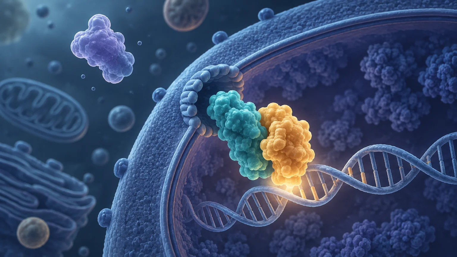
TLDR: The NF-kB p50 p65 heterodimer is a transcription-factor complex formed by p50, a processed product of the NFKB1 precursor p105, and p65, also called RelA. Their conserved Rel homology domains support dimerization, DNA recognition, inhibitor binding, and localization control. In the canonical pathway, degradation of IκBα permits nuclear accumulation of NF-κB. DNA binding involves both subunits, but much of the complex’s classical transcriptional activation capacity comes from the C-terminal transactivation domain of p65. Nuclear p65 alone does not prove p50/p65 formation, binding at a particular gene, or transcriptional activation.
The important point is that “NF-κB activation” is not one molecular event. It can refer to inhibitor degradation, nuclear localization, DNA binding, chromatin occupancy, transcription, or a downstream phenotype. Understanding how the NF-kB p50 p65 heterodimer works makes it easier to choose an assay and avoid treating one readout as proof of every later step.
What is the NF-kB p50 p65 heterodimer?
NF-κB is a family of related transcription factors rather than a single protein. The classic p50/p65 complex contains one p50 subunit and one p65 subunit. p65 is another name for RelA and is encoded by RELA. p50 is generated through processing of the larger NFKB1-encoded p105 precursor. It is therefore more precise to say that NFKB1 encodes p105 and gives rise to p50 than to treat p50 and full-length p105 as interchangeable proteins.
The p50/RelA combination is a central dimer in canonical NF-κB signaling, but it is not the only possible NF-κB complex. Family members can form multiple homo- and heterodimers, and the predominant complex can vary with cell type, stimulus, concentration, and time after stimulation. A generic signal for p65 or NF-κB activity should not automatically be interpreted as proof that p50/p65 is the only relevant dimer.
| Subunit | Gene or precursor relationship | Key functional feature |
|---|---|---|
| p50 | Produced from the NFKB1 p105 precursor | Contributes to dimerization and κB-site DNA recognition but lacks a p65-like classical C-terminal transactivation domain |
| p65 | Also called RelA; encoded by RELA | Contributes to DNA recognition and carries a C-terminal transactivation domain |
| p50/p65 | Heterodimer containing one of each subunit | Combines sequence recognition with strong context-dependent transcriptional activation potential |
The Rel homology domain organizes dimerization and DNA binding
Both p50 and p65 contain a conserved Rel homology domain, commonly abbreviated RHD. This region is multifunctional: it contributes to dimer formation, recognition of κB DNA elements, interactions with IκB inhibitory proteins, and regulated nuclear localization. These functions are structurally connected, which helps explain why NF-κB behavior depends on the state of the entire complex rather than on one isolated sequence motif.
At the dimerization step, complementary surfaces within the RHD allow p50 and p65 to associate. The resulting heterodimer then presents DNA-contacting surfaces from both subunits. The dimer should therefore be understood as an integrated DNA-binding assembly, not as p50 merely carrying p65 to DNA or p65 acting independently of p50.
A useful structural reference is the crystal structure of the DNA-bound p50/p65 heterodimer. That work resolved the dimer interface and showed p50 and p65 contacting distinct portions of an immunoglobulin light-chain enhancer κB element. The structure provides direct evidence for how the two subunits cooperate at a specific DNA site, although one crystallized complex cannot represent every chromatin context encountered in a cell.
How p50/p65 recognizes κB DNA elements
NF-κB dimers bind regulatory DNA elements generally called κB sites, which occur in promoters and enhancers. Recognition is sequence-sensitive, but a consensus motif is only a starting point. Different κB sequences can influence affinity, conformation, cofactor use, and regulatory output. Chromatin accessibility, neighboring regulatory elements, protein concentrations, and cell state further determine whether an apparently suitable motif is occupied in vivo.
One biochemical study measured an affinity of roughly 10 nM for p50/p65 binding to a particular immunoglobulin κ enhancer κB site. That value is useful for understanding high-affinity binding under the reported assay conditions, but it is not a universal dissociation constant for NF-κB. Changing the sequence, dimer composition, buffer, competitor DNA, or other assay conditions can change the measured interaction.
This distinction matters when interpreting genomic sequence scans. Finding a κB-like motif establishes that a locus could be compatible with NF-κB recognition. It does not establish that p50/p65 occupies the site in the relevant cells, under the relevant stimulus, or at the relevant time. Direct occupancy measurements and transcriptional evidence are needed to move beyond motif-based prediction.
Why the p65 transactivation domain matters
DNA binding and transcriptional activation are related but separable functions. The p65 subunit contains a C-terminal transactivation domain that can support recruitment and regulation of transcriptional machinery and cofactors. p50 lacks the corresponding classical C-terminal transactivation domain. This asymmetry helps explain why p50/p65 is commonly associated with transcriptional activation: p50 contributes to the DNA-bound complex while p65 supplies substantial transactivation capacity.
That explanation should not be reduced to “p65 always activates and p50 always represses.” Regulatory output depends on the DNA site, chromatin environment, post-translational state, cofactors, dimer partner, and stimulus history. The absence of a p65-like transactivation domain makes p50/p50 mechanistically different from p50/p65, but it does not assign one unavoidable effect to every p50-containing complex.
Even confirmed p50/p65 binding does not guarantee increased expression of a nearby gene. Occupancy can be nonproductive, transient, or dependent on missing cofactors. Conversely, changes in gene expression after pathway stimulation may be indirect. A strong mechanistic chain connects dimer identity, locus occupancy, transcriptional change, and—when relevant—functional outcome using complementary experiments.
IκBα, IKK, and regulated nuclear localization
In many resting cells, p50/p65 is associated with IκBα and retained predominantly outside the nucleus. In the canonical pathway, appropriate upstream signals activate the IκB kinase complex, or IKK. IKK-dependent phosphorylation of IκBα promotes its ubiquitination and proteasomal degradation, releasing NF-κB from inhibition and permitting nuclear accumulation.
The localization mechanism is more precise than the shorthand statement that NF-κB simply “switches on and enters the nucleus.” IκBα can mask the nuclear localization signal of p65, making localization a property of the inhibitor-bound assembly. Removing the inhibitor changes which localization information is available to the cellular transport machinery.
NF-κB signaling also changes over time. Depending on the stimulus and cellular context, activity can be transient, sustained, or oscillatory. Feedback is important: NF-κB can promote expression of inhibitory components, including IκBα, which can help terminate or reshape the response. A single endpoint can therefore miss a peak, a delayed response, or repeated cycles of nuclear localization.
p50/p65 compared with p50/p50 and other dimers
Subunit composition changes what an NF-κB DNA-binding signal means. A p50/p65 heterodimer contains the p65 transactivation domain, whereas a p50/p50 homodimer does not. p50/p50 can produce different regulatory outcomes depending on associated cofactors and sequence context. Other NF-κB family combinations add further diversity.
- p50/p65: combines the DNA-binding properties of both RHDs with the C-terminal transactivation capacity of p65.
- p50/p50: lacks a p65-like transactivation domain and may behave differently depending on cofactors and chromatin context.
- Other NF-κB dimers: can differ in stimulus coupling, preferred regulatory sites, localization, kinetics, and transcriptional effects.
Consequently, an electrophoretic mobility shift or reporter response labeled broadly as “NF-κB activity” may combine contributions from several dimers. Establishing p50/p65 specifically requires composition-sensitive evidence rather than relying only on the pathway name.
What common NF-κB experiments can—and cannot—show
The most informative assay depends on which mechanistic step is being tested. No single standard readout resolves localization, dimer composition, endogenous DNA occupancy, transcription, and phenotype at once.
Immunofluorescence and cellular fractionation
Immunofluorescence can show redistribution of p65 toward the nucleus, provided image acquisition, segmentation, antibody specificity, and nuclear boundaries are adequately controlled. Biochemical fractionation can compare nuclear and cytoplasmic pools, but interpretation depends on demonstrating fraction purity and avoiding leakage or cross-contamination.
Either method can support nuclear accumulation. Neither method alone proves that nuclear p65 is paired with p50, bound to a particular κB element, or activating transcription. Nuclear p65 is therefore evidence consistent with pathway activation, not a complete mechanism.
EMSA and composition-sensitive variants
An electrophoretic mobility shift assay, or EMSA, tests whether proteins in a sample form complexes with a supplied DNA probe. Competition with unlabeled probe can assess sequence-dependent binding, while antibody-dependent supershift or disruption can help identify subunits. An ordinary shifted band, however, does not establish endogenous chromatin occupancy and may not uniquely identify p50/p65 without composition-sensitive controls.
ChIP and related occupancy assays
Chromatin immunoprecipitation can test whether p65 or p50 is enriched at a selected genomic region in cells. Appropriate input, negative regions, antibody validation, and biological controls are essential. Separate p50 and p65 enrichment at the same region is consistent with co-occupancy, but ordinary ChIP does not necessarily prove that both proteins were present in the same molecular complex at the same moment.
Reporter assays and endogenous transcription
A κB-responsive reporter measures the output of an engineered regulatory construct. It is useful for testing pathway-responsive transcription under controlled conditions, but it may not reproduce endogenous chromatin or identify which NF-κB dimer generated the signal. Reporter activity can also be affected by cell health, transfection efficiency, normalization strategy, and signaling pathways that alter the reporter system indirectly.
For endogenous output, RNA measurements can test whether candidate transcripts change after stimulation or perturbation. Protein abundance and functional assays may then be needed because transcript change is not equivalent to protein production or biological effect.
A practical evidence ladder
- Test pathway initiation with appropriate measurements of IKK activity or IκBα modification and loss.
- Measure the timing and magnitude of p65 and p50 nuclear accumulation.
- Use composition-sensitive DNA-binding or protein-interaction methods to test p50/p65 formation.
- Measure occupancy at relevant endogenous loci rather than relying only on motif prediction.
- Test endogenous RNA and, where meaningful, protein output.
- Use perturbation, rescue, or loss-of-function designs to connect the complex to the proposed phenotype.
Not every project requires all six steps. The ladder instead clarifies the strongest conclusion available from the evidence collected. For example, nuclear p65 supports localization; ChIP enrichment supports locus occupancy; and altered target RNA supports transcriptional output. None should silently substitute for the others.
Disease research without therapeutic overinterpretation
NF-κB signaling is widely investigated in immune signaling, inflammation, stress responses, cell survival, and cancer biology. Its biological importance does not mean that every increase in nuclear p65 is disease-causing or that every inhibitor of an NF-κB readout will be therapeutically useful.
Biochemical experiments can establish molecular interactions. Cultured-cell studies can identify context-dependent signaling and phenotypes. Animal models can test integrated responses in a particular model organism. None of these evidence levels alone establishes clinical efficacy, safety, an appropriate human dose, or the value of targeting NF-κB in a specific patient population.
The pathway also has broad, context-dependent functions, so pathway suppression and beneficial disease modification cannot be treated as synonyms. A responsible disease claim must identify the experimental model, stimulus, intervention, timing, measured endpoint, and causal evidence. A broad reporter decrease or reduced p65 nuclear signal is mechanistic evidence at one level—not treatment guidance.
Key takeaway
The p50/p65 NF-κB heterodimer is best understood as a regulated molecular assembly. p50 comes from processing of NFKB1 p105; p65 is RelA; their Rel homology domains support dimerization and κB-site recognition; and the p65 C-terminal domain supplies important transactivation capacity. IκBα controls access to the nucleus, while IKK-dependent signaling can release that restraint.
For experimental interpretation, first name the step actually measured: inhibitor degradation, nuclear localization, dimer composition, DNA binding, endogenous occupancy, transcription, or phenotype. Then choose a complementary assay for the next step in the proposed mechanism. That discipline turns a general statement about “NF-κB activation” into a testable and appropriately bounded conclusion.
References
- The NF-κB Family of Transcription Factors and Its Regulation – PMC
- Structural motifs involved in ubiquitin-mediated processing of the NF-kappaB precursor p105: roles of the glycine-rich region and a downstream ubiquitination domain.
- NF-κB signaling – PubMed
- Structural studies of NF-κB signaling – PMC
- Crystal structure of p50/p65 heterodimer of transcription factor NF-kappaB bound to DNA – PubMed
- Mechanism of kappa B DNA binding by Rel/NF-kappa B dimers – PubMed
- Independent modes of transcriptional activation by the p50 and p65 subunits of NF-kappa B.
- Crosstalk via the NF-κB Signaling System – PMC