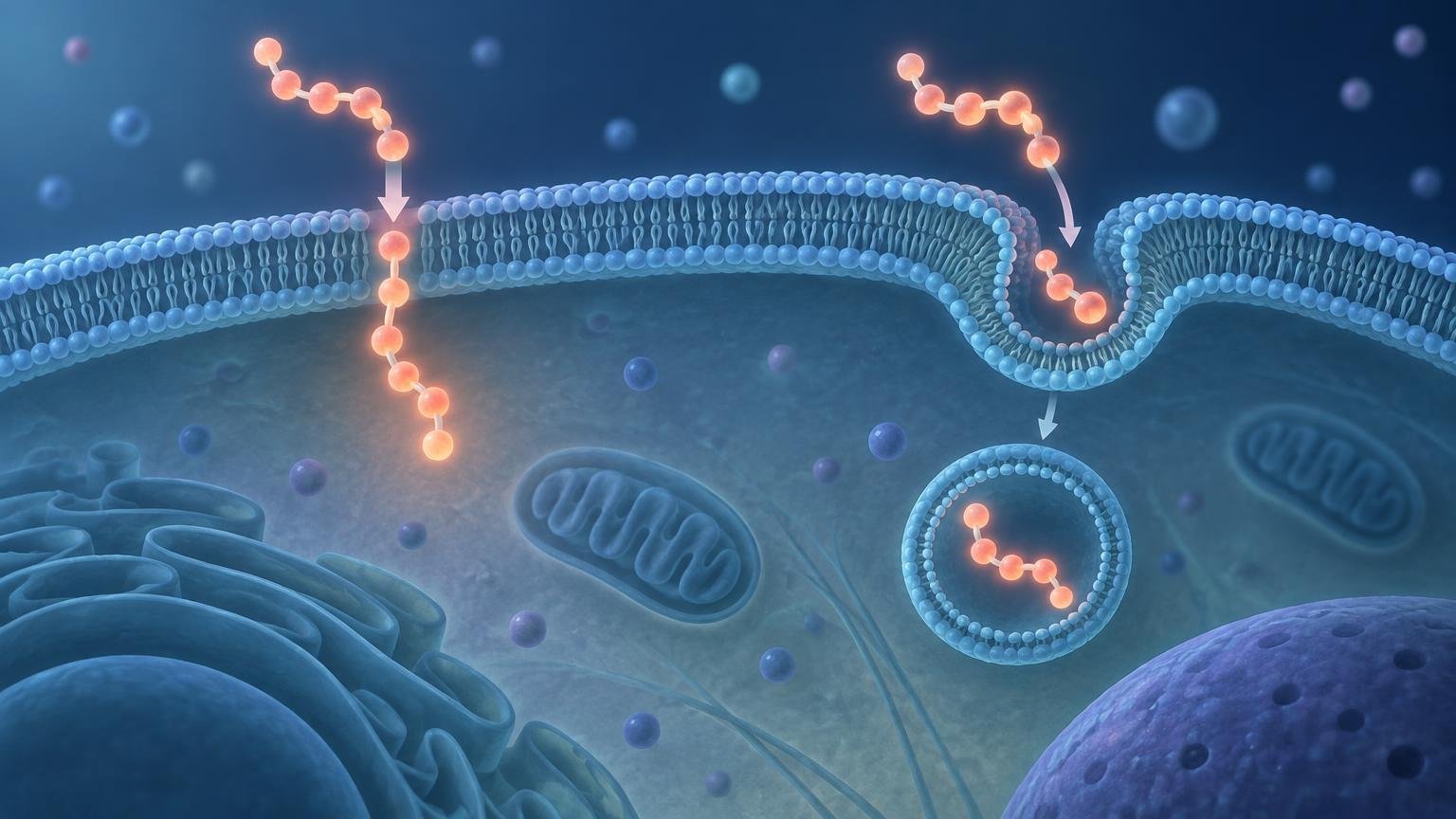
TLDR: The cell penetrating peptide direct translocation vs endocytosis question does not have one answer for every peptide. A CPP may cross the plasma membrane without initial vesicular sequestration, enter through an endocytic pathway, or use both routes under different conditions. The scientifically useful question is whether a defined CPP–cargo construct reaches the required intracellular compartment without causing membrane damage—and whether the experiment actually distinguishes surface binding, vesicular uptake, and functional cytosolic delivery.
Reports supporting both routes are not necessarily contradictory. CPP sequence, cargo architecture, concentration, cell type, membrane composition, exposure time, and measurement method can all shift the apparent balance between direct entry and endocytosis. Quantitative studies therefore argue against treating uptake as a universal either-or property of the CPP sequence alone.
What direct translocation and endocytosis mean
Direct translocation is an outcome-level description: the peptide crosses the plasma membrane without first being enclosed in a conventional endocytic vesicle. If the cargo remains attached and functional, this route could provide relatively immediate access to the cytosol. The term does not identify a single molecular mechanism. Membrane deformation, transient defects, lipid rearrangement, and vesicle-like budding followed by collapse have all been proposed in particular experimental systems.
Endocytosis begins differently. The plasma membrane bends around extracellular material and encloses it in an intracellular vesicle. CPPs have been associated with multiple endocytic processes, including macropinocytosis and other energy-dependent pathways. The route can depend on how a peptide interacts with membrane lipids, glycans, receptors, or other cell-surface components.
| Feature | Direct translocation | Endocytosis-mediated uptake |
|---|---|---|
| Initial membrane event | Crossing without initial sequestration in a conventional endocytic vesicle | Membrane enclosure followed by formation of an intracellular vesicle |
| Early localization expected | Cytosolic availability may occur rapidly if crossing is productive | Punctate or vesicular localization is commonly expected |
| Major interpretation problem | Membrane disruption can resemble productive entry | Internalization can be mistaken for cytosolic delivery |
| Additional barrier | Cargo release and correct localization may still be required | Endosomal escape, cargo release, and correct localization may all be required |
| Useful evidence | Live-cell entry with membrane-integrity and functional-delivery controls | Colocalization, pathway perturbation, trafficking analysis, escape measurements, and functional delivery |
These categories are useful, but biology does not have to respect them as mutually exclusive labels. A cell population may contain vesicular and diffuse peptide at the same time. Individual cells may differ, and the dominant route may change during an experiment.
Why the route matters for useful intracellular delivery
Total cellular association is not the same as cytosolic availability. A fluorescent CPP can bind strongly to the cell surface, enter vesicles, or accumulate in endosomes while producing a large total signal. None of those observations alone demonstrates that an attached cargo is free in the cytosol.
This creates a sequence of distinct experimental questions: Did the construct bind the cell? Was it internalized? Is it trapped in an endosome? Did it escape? Was the cargo released if release is required? Did it reach and engage its intracellular target? Did that engagement produce the expected functional response? A methods-focused discussion of cell-surface binding versus true CPP internalization explains why these stages need separate measurements.
Endocytosis can be an efficient way to concentrate material inside a cell, but it also creates the endosomal-escape problem. A cargo intended to inhibit a cytosolic enzyme, bind a transcription factor, or reach the nucleus generally cannot complete that task while confined within an intact endosome. Conversely, a diffuse cytosolic signal is not automatically desirable if it results from nonspecific plasma-membrane damage.
Evidence for endocytosis-mediated CPP uptake
Arginine-rich CPPs can interact with negatively charged components at the cell surface. In one studied system, membrane-associated proteoglycans were important for actin organization and macropinocytic uptake. Macropinocytosis involves actin-dependent membrane remodeling and formation of comparatively large vesicles that take up extracellular fluid and associated material.
Evidence consistent with endocytosis may include punctate intracellular localization, overlap with endosomal markers, reduced uptake after perturbing an implicated pathway, temperature or energy dependence, and time-dependent trafficking through vesicular compartments. No single observation is conclusive. A pathway inhibitor may have off-target effects, while low temperature can alter membrane behavior and cellular metabolism more broadly than the pathway being investigated.
Vesicular fluorescence establishes localization more directly than total fluorescence, but it still does not quantify how much material has escaped. A small cytosolic fraction can sometimes drive a substantial functional effect, whereas a large endosomal pool may remain biologically unavailable. Localization and function should therefore be measured independently.
Evidence for direct or endocytosis-independent entry
Direct entry cannot be dismissed simply because endocytosis is common. Live-cell work has reported endocytosis-independent entry of arginine-rich peptides under defined conditions, showing that not all observed CPP internalization must proceed through endocytosis. Earlier mechanistic debate was shaped in part by a landmark live-cell reevaluation of CPP uptake, which demonstrated how experimental handling could change the apparent localization pattern.
Direct-entry evidence is strongest when movement across the plasma membrane is observed in living cells, persists after surface-associated signal is removed or quenched, occurs without the expected vesicular intermediates, and is accompanied by a cytosolic functional readout. Membrane-integrity controls are essential. Leakage of normally excluded dyes, loss of cytosolic contents, or rapid toxicity would suggest membrane injury rather than controlled delivery.
Proposed direct-translocation mechanisms
There is no universally accepted molecular mechanism covering every CPP. Arginine-rich peptides have been reported to induce transient focal membrane deformation associated with direct penetration in a tested system. Other models invoke local membrane thinning, transient defects, or rearranged lipid structures that reduce the energetic cost of moving a charged peptide through the bilayer.
A 2024 study proposed another model in which R9 and cyclic CPPs induce plasma-membrane vesicle budding followed by collapse, producing translocation in the tested cells. This is an experimentally supported proposal for those systems, not a field-wide resolution. Different observations may reflect distinct molecular events, or different views of a dynamic membrane-remodeling process.
Why a CPP can appear to change routes
Peptide sequence and chemistry
Charge distribution, arginine or lysine content, hydrophobicity, amphipathicity, conformation, and multivalency can change how a CPP interacts with membranes and cell-surface molecules. Even closely related sequences may differ in surface binding, membrane remodeling, endocytic recruitment, and intracellular trafficking. Quantitative studies have found that sequence and cell-surface components can affect the balance between translocation and endocytosis.
Cargo size, linkage, and valency
A CPP should not be evaluated independently of its cargo. Attaching a protein, oligonucleotide, nanoparticle, or multivalent scaffold changes the size, charge, shape, and flexibility of the complete construct. It may also hide CPP residues or create new interactions at the membrane.
In tumor-cell models, one study reported rapid direct translocation alongside endocytic uptake for small CPP cargoes, while larger linked components tended toward slower, energy-dependent macropinocytic entry. This result supports cargo-dependent behavior in that experimental system; it does not define a universal size cutoff. For a broader design framework, see how cargo size changes CPP delivery.
Concentration and exposure conditions
Concentration can change the density of peptide at the membrane. Below one range, uptake may be dominated by binding and endocytosis; at higher local density, cooperative membrane remodeling may become more apparent. However, there is no universal concentration threshold or guaranteed direction of change across CPPs. High concentrations also increase the need to test membrane disruption, aggregation, and cytotoxicity.
Cell type and membrane composition
Cells differ in membrane lipid composition, surface proteoglycans, endocytic activity, metabolic state, and membrane-repair capacity. Those differences can change both peptide binding and the response to that binding. A route inferred in one immortalized cell line should not be assumed to dominate in primary cells, another tissue lineage, or an in vivo environment.
The measurement problem in cell penetrating peptide direct translocation vs endocytosis
The assay can create the apparent mechanism. Fixed-cell imaging was historically a major source of confusion because fixation can redistribute surface-associated or vesicular peptides, creating diffuse intracellular patterns that resemble direct cytosolic entry. Live-cell reevaluations documented this risk for CPPs and protein-transduction domains.
Fluorescent labels introduce additional variables. A fluorophore changes the construct's mass, hydrophobicity, and charge; its signal can also depend on pH, local concentration, cleavage, or degradation. Flow cytometry measures signal associated with individual cells but does not inherently identify its subcellular location. Fluorescence spectroscopy can quantify bulk signal yet has a similar localization problem. Comparative work using fluorescence methods and mass-spectrometry quantification illustrates why the analytical method can influence the apparent uptake result.
Microscopy supplies spatial information, but diffuse signal can be difficult to distinguish from out-of-focus vesicles or redistributed material. Punctate signal supports vesicular localization but does not show that every molecule is trapped. A convincing route assignment normally requires orthogonal methods rather than a single image or total-uptake number.
A practical checklist for evaluating a CPP uptake study
- Define the complete construct. Record the CPP sequence, cargo, linker, label, charge, size, and whether cargo release is expected.
- Use living cells for the primary localization experiment. Treat fixed-cell images as supporting evidence unless fixation artifacts have been specifically controlled.
- Separate extracellular and surface-bound signal from internal signal through validated washing, protease treatment, quenching, or another appropriate method.
- Measure membrane integrity. Include leakage, dye-exclusion, viability, and morphology controls at the same concentration and exposure used for delivery.
- Track vesicular localization. Use suitable endosomal markers and time-resolved imaging rather than interpreting punctate fluorescence without compartment identification.
- Test pathway dependence cautiously. Combine pharmacological perturbations with genetic or orthogonal approaches where practical, because inhibitors and temperature shifts are rarely pathway-specific.
- Use more than one quantification method. Imaging, flow cytometry, biochemical fractionation, mass spectrometry, or other approaches answer different parts of the question.
- Verify fraction purity if biochemical fractionation is used. Contamination between membrane, endosomal, and cytosolic fractions can create false localization conclusions.
- Include a functional cytosolic-delivery assay. A cargo-dependent response that requires access to a cytosolic or nuclear target is more informative than total fluorescence alone.
- Demonstrate cargo integrity and activity. Uptake of a label or peptide fragment does not establish delivery of an intact, functional conjugate.
- Test multiple concentrations, exposure times, and relevant cell types. Mechanistic conclusions drawn from one condition should remain condition-specific.
The strongest design connects localization to function without using function alone as proof of route. A downstream phenotype could arise from surface signaling, stress, membrane damage, or an off-target effect. Ideally, target engagement, cargo dependence, and rescue or loss-of-function controls connect intracellular availability to the observed response.
What cultured-cell uptake does not establish
Efficient uptake in cultured cells does not establish tissue penetration, selective delivery, acceptable exposure, safety, or therapeutic efficacy. A construct must also survive biological fluids, reach the relevant tissue, distribute within that tissue, enter the intended cells, and avoid unacceptable off-target effects. These are separate experimental questions.
The evidence ladder matters: biochemical membrane models, cultured cells, animal models, and human studies answer progressively different questions. The distinctions are discussed further in how biotechnology evidence changes from cell culture to human trials. CPP uptake studies can support a delivery mechanism under defined conditions, but they do not by themselves demonstrate disease-treatment utility.
Conclusion
Direct translocation and endocytosis should be treated as context-dependent routes, not competing universal explanations. The same CPP may use both, and changing its cargo, concentration, cell system, or assay can change which route appears dominant.
The most useful next step is to define the desired delivery endpoint before choosing an assay. If the cargo needs cytosolic access, measure cytosolic availability and function—not merely cellular fluorescence. If endocytosis dominates, quantify escape rather than assuming it. If direct entry is proposed, show that it occurs in living cells without membrane damage. That evidence-based sequence turns a route label into a meaningful intracellular-delivery conclusion.
References
- Cell-penetrating peptides. A reevaluation of the mechanism of cellular uptake – PubMed
- Translocation and endocytosis for cell-penetrating peptide internalization – PubMed
- Quantitative fluorescence spectroscopy and flow cytometry analyses of cell-penetrating peptides internalization pathways: optimization, pitfalls, comparison with mass spectrometry quantification – PubMed
- Cell entry of arginine-rich peptides is independent of endocytosis.
- Interaction of arginine-rich peptides with membrane-associated proteoglycans is crucial for induction of actin organization and macropinocytosis – PubMed
- Transient Focal Membrane Deformation Induced by Arginine-rich Peptides Leads to Their Direct Penetration into Cells – PMC
- Cell-Penetrating Peptides Translocate across the Plasma Membrane by Inducing Vesicle Budding and Collapse – PubMed
- Distinct transduction modes of arginine-rich cell-penetrating peptides for cargo delivery into tumor cells.
- Cell surface adherence and endocytosis of protein transduction domains – PubMed